New Tools for Bladder Research
Over the last 10 years, new microscopy tools have aimed at two things: higher magnification and better resolution. This is great for imaging cells and tissue slices, but confocal/multiphoton microscopy becomes pretty useless when you’re imaging a spherical hollow organ like the bladder. So, we sought to change that.
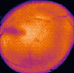
So how do you take meaningful pictures of the entirety of a hollow sphere? Well, as usual — the simplest answer is the right one. Using good-old-fashioned wide-field microscopy techniques and genetically-encoded calcium biosensors, we are able to visualize calcium signals traveling through a network of cells in the bladder wall (presumably interstitial cells). The function of these waves is unknown, but we’re planning to figure that out.

But what’s happening on the other side of the bladder? or the other other side? Enter the Pentaplanar Reflected Image Macroscopy System — or PRIM for short. This device (designed by our lab and patent-pending) allows us, for the first time, to measure micromotions across the entirety of the bladder wall in a single video. Coupled with our calcium biosensor mice and afferent nerve recordings, this system is at the forefront of integrated bladder physiology research.
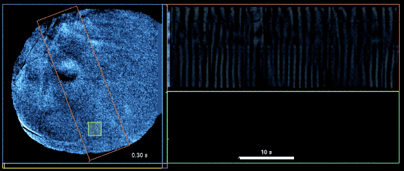
What do we do with these movies, now that we have them? Since nothing like this has been recorded before, we needed to develop new software systems to bring these images to new heights. With state-of-the-art stabilization and differentiation algorithms, we are able to map changes in location and intensity of calcium signals, as well as isolate the dynamic calcium changes from background noise.